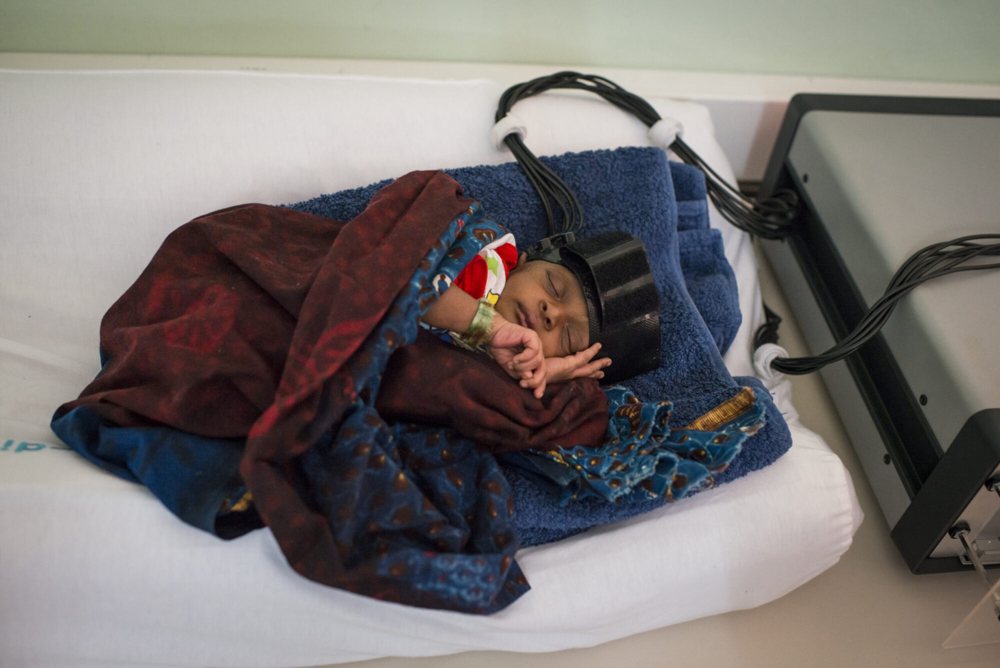
The lateral row may require a visit to your school gymnasium, which is a good excuse to get your pump on after you do your experiments. Muscle fatigue occurs in everyday life and can be described as a feeling or sensation of weakness or muscle pain. Static and dynamic myoelectric measures of shoulder muscle fatigue during intermittent dynamic exertions of low to moderate intensity. Lots of research has been done into looking at this difference (example), and it has been found (by us too!) Tucker, Project management: J.M. Clipboard, Search History, and several other advanced features are temporarily unavailable. Beginning at age 50, muscle mass decreases at an annual rate of 1% to 2%, and a decline in muscle strength of 1.5% to 3% per year is documented.22 Further research is needed to understand muscular physiological changes in HAD and their influence on muscle fatigue. With the exception of the vastus lateralis during a dynamic contraction in healthy younger and hospitalized patients, there was a statistically significant difference between Fmed at initiation and termination of contraction, indicating that subjects muscles did truly fatigue. Eur J Appl Physiol. By squaring it! Write down the length of time the subject was contracting as shown by the EMG, and save the recording with identifiable notation, such as "Muscle Fatigue - Woman 1 - total time 118s." If a nerve cannot maintain a high intensity spiking signal, secreting neurotransmitters constantly, it experiences "synaptic fatigue," where the nerve is no longer able to stimulate the muscle fibers it innervates. The Kruskal-Wallis test showed that there were statistically significant differences in TTTF between groups after an isometric contraction (P=.03) and a dynamic contraction (P=.04) (Tab. Functional outcomes with early rehabilitation in the acute care setting have improved; however, an improved understanding of muscle fatigue using surface electromyography (sEMG) is warranted to better guide patient-centered exercise prescription. A limitation of the study was the small sample size of patients who were hospitalized without matched controls. Epub 2015 Jul 13. Since our experiment started at time = 0, 0 will be the second x variable. IN , Schlinder-Delap B, Hunter S. Medrinal 2021 Feb 3;21(4):1024. doi: 10.3390/s21041024. . This was done by nonparametric paired t tests with Wilcoxon post hoc analysis. Calculating the average rates of fatigue, the women had an average rate -0.06 mV/s, and the men came in at -0.11 mV/s. Full Document. On average, during contractions of 10% MVC no EMG changes were detected. endobj 13 0 obj Now let us consider the case of Robert. Now calculate the slope, or rate of fatigue, for all your subjects. Additionally, the study of muscle fatigue, using these measurements, in an acutely ill patient population adds to the current limited existing knowledge. Course Hero is not sponsored or endorsed by any college or university. Record in L play to continue 45 s 60 s 75 s At 60 seconds, m count number of units. not necessarily, endurance isn't really about strength. Record in La This site needs JavaScript to work properly. Please enable it to take advantage of the complete set of features! TIME TO FATIGUE), Lab Data Training. <>/Border[0 0 0]/P 3 0 R>> You can contribute to the world's scientific knowledge! sEMG is a lab quantitative technique that was found to be safe and feasible to assess muscle fatigue in the acute care environment. All testing was performed with the subjects in a semireclined position and conducted by the same tester (J.M.S.). We can go full circle here and practice calculating the trend line, or line of best fit. Time NN 60 5 75 5 19 1 Time to Fatigue (s) This problem has been solved! At 75 seconds, License. View Surface EMG was measured from 14 shoulder muscles while participants performed simulated, repetitive work tasks until exhaustion. Our preliminary data thus suggests that women may have better muscle endurance, fatiguing at a slower rate! Method: The https:// ensures that you are connecting to the 2 0 obj The site is secure. Each time a muscle fiber is activated, an action potential is conducted from the nerve, along the muscle fiber, and into the fiber. Suriyaarachchi , Fan E, Brower R, Needham D. Kortebein Bethesda, MD 20894, Web Policies J 1959 Aug;38:148-58 This was an observational cohort study between healthy younger participants, healthy older participants, and patients who were hospitalized. 2021 Oct 22;21(21):7014. doi: 10.3390/s21217014. The authors acknowledge the following individuals: Christina Carpenter, PT, DPT; Ning Gong, PhD; Sheena MacFarland, PT, DPT; Jenna Tosta, PT, DPT; and Josh Tucker, BS. Record in La play to continue 08 15 s 30 s At 45 seconds, me count number of a units. Although it is well known that patients muscles during hospitalization develop atrophy and weakness,11 little is known about peripheral muscle fatigability in this patient population. Time is our x variable and RMS is our y variable. For each recording you thus have three measurements 1) total time in seconds - Total Time(s), 2) RMS of the first five seconds - RMS Begin, and 3) RMS of the last five seconds - RMS End. Our favorites are the bicep lift (which uses the biceps), the deltoid lift (which uses the deltoid), and the lateral row (which uses the latissimus dorsi). In one experiment, intermittent static arm pulling (triceps) (10 s contraction and 5 s rest, average work load 14% and 10% MVC) was performed for 7 h. The endurance time for the sustained contractions was around one hour for 10% MVC, and it was shown--all in all--that the concept of "indefinite" endurance times at contractions below 15-20% MVC cannot be maintained. and transmitted securely. De Jonghe Time to fatigue Skrzat, S.J. Muscle fatigue is defined as any exercise-induced reduction in the ability of the muscle to produce force or power.12 Fatigue, also known as time-to-task-failure (TTTF), is the time from the initiation of a force-generating contraction to termination of contraction and is commonly measured using surface electromyography (sEMG). the number of motor units that are active at regular intervals. The more values, the more compelling the results! Representative median frequency electromyographic (EMG) data of the first 5s (A) and the last 5s (B) of an isometric contraction in a hospitalized patients rectus femoris. A new framework for prediction of task failure using EMG amplitude data alone is presented. play to continue The PubMed wordmark and PubMed logo are registered trademarks of the U.S. Department of Health and Human Services (HHS). Look at the RMS value displayed on the screen and write it down. Note, this is only an issue if you have our normal, Choose a weight load that the subject is comfortable maintaining between 15-120 seconds before load fatigue is too high. The Backyard Brains Spike Recorder program allows you to visualize and save the data on your computer when doing experiments. Sensors (Basel). Now that we've got our values, let's chart them out to make them easier to manage. strength. Results: The depressed gas pedal, like the electric stimulation, forces the engine to run strong. 60 s 24 2 An IMU (Shimmer3 w/450mAH Battery; Shimmer, Dublin, Ireland), attached approximately 1 inch (2.5 cm) lateral and distal to the inferior angle of the patella, was used to determine leg position during the active contraction. PMC 15 s >VPO];lS4IJIM ,,2,v,0|*MrC3>G[Nc-%*A+|VkmnlUtS}iYGVgVSX0-iw4'\x4o_fdWux8l|-~'5v683c~b}r+:K`FY|/LST#lN5Jcv5-oG295bY_7nt\EGlua>N~Zvz~c{wsOZqFEwxwd0at.m?RyJnz$yh.F}W6FIegrrlth__276z^[^jiVO[S%Wz. Patients who were hospitalized were screened by J.M.S. , Bronco A, Arrigoni Marocco S, et al. The electrical signal is emitted by the muscle fiber during each activation and subsequently detected by the sEMG sensor.12 sEMG sensors are wireless and compact, allowing assessment of muscle activity in a multitude of environments with minimal impact on typical movement patterns. How to Measure Myoelectric signals are formed by physiological variations in the state of muscle fiber membranes." (Basmajian, J.V. Are they stronger? 2018 Apr;39:58-69. doi: 10.1016/j.jelekin.2018.01.001. Concept/idea/research design: J.M. Maybe! not necessarily, endurance isn't really about strength. Bookshelf Epub 2007 Jul 20. Record in The https:// ensures that you are connecting to the Next, we want to adjust the gain. P Once you can, have your subject relax a couple of minutes. HO=healthy older; HP=hospitalized patient; HY=healthy younger; MRC Score=Medical Research Council Score of Strength; MVIC=maximal voluntary isometric contraction. Observe the EMG screen for the maximum force, pause the screen. PHASES, ec facilisis. . To transform the EMG signal in the time-domain to the frequency-domain, a Fourier transform of the autocorrelation function of the EMG signal is employed to provide the power spectrum (PS) or the power spectral density (PSD). Question: ELECTROMYOGRAPHY. Patients in the hospital are susceptible to development of HAD. National Library of Medicine Pellentesque dapibus efficitur laoreet. 5. SHOW LABELS For this experiment, you may be interested in comparing young versus old subjects, boys versus girls, or maybe you want to compare people who have a high-weight max bench press versus a low-weight max. We can solve for the rate of fatigue by plugging into the formula for the slope above. 4 B Scroll to marker labeled "Non dominant arm: Continued clench at maximum 7 0 obj [XR-Wb`lq8k%0gjhExbU*q-x5"5xvj[P;>cEcea~S.Qd*~e cCl(%NKI"!~B"cJNaGkpM - qYApMH&u"q$Od^G /v`FRx~ZHJr0h9lC8S4WZe`RjXf*A9N!w,ME:i0"l9YaK`CJ`lIJYGc&Ks. HWe will get there, hold on. People with Multiple Sclerosis typically exhibit greater levels of exercise-induced fatigue compared to healthy individuals. Now we take the "sum of the squares" of the signal and divide it by the number of samples we took to get our mean (or average) value. GO TO PH (10 s contraction and 5 s rest, average work load 14% and 10% MVC) was performed for 7 h. The endurance time for the sustained contractions . <>/Border[0 0 0]/P 3 0 R>> units. Before you begin, make sure you have the Backyard Brains Spike Recorder installed on your computer/smartphone/tablet. Calculating the average rates of fatigue, the women had an average rate -0.06 mV/s, and the men came in at -0.11 mV/s. Both force and mot. Our secondary analyses of Fmed by group showed that there was a statistically significant change in Fmed between the first 5seconds and last 5seconds of both isometric and dynamic muscle contractions, with the exception of the Fmed of the healthy younger participants and hospitalized patients VL during the dynamic contraction (Tab. That was the beginning. Bookshelf Record in La play to continue 08 15 s 30 s At 45 seconds, me count number of a units. 75 s 19 1, Copyright 2023 StudeerSnel B.V., Keizersgracht 424, 1016 GC Amsterdam, KVK: 56829787, BTW: NL852321363B01, Give Me Liberty! Sample graphics can be found in Appendices 13. After 5% MVC sustained contractions for one hour a 12% reduction in MVC was seen, and significant increases in EMG amplitude and decreases in the mean spectral frequency of the EMG-power spectrum were found. The authors developed a function to quantify fatigue in multiple shoulder muscles by generating a single score using relative changes in EMG amplitude and frequency over time. Maybe! Zanni Experts are tested by Chegg as specialists in their subject area. 30 s 2008 Dec;18(6):1006-19. doi: 10.1016/j.jelekin.2007.03.012. Outliers were removed if they were more than 2 SDs from the mean. sEMG is a lab quantitative technique that was found to be safe and feasible to assess muscle fatigue, with a protocol that yielded similar results to previously published literature. The mean frequency and median frequency of the EMG signals were extracted as dataset features. Studying muscle fatigue can assist the physical therapist in implementing a patient-centered exercise prescription. electromyography; fatigue; muscles; quantification; shoulder. The periodogram function was used to output a frequency vector and power spectral density estimate of the signal (PSD_EMG) using the sEMG signal and sampling frequency as inputs. 1985 Oct;125(2):265-75 The site is secure. The authors completed the ICMJE Form for Disclosure of Potential Conflicts of Interest and reported no conflicts of interest. What can the slope of the trend line teach you about muscle fatigue? Please check for further notifications by email. Why, it will be the amplitude of our EMG! This is the most purely muscle physiology variation of fatigue. Further research is needed to better understand how to integrate sEMG findings into the development of patient-centered exercise prescriptions. The function was evaluated through changes in MMFS over time: first (31.8 14.6), middle (47.6 25.3), last (58.6 35.5) reference exertions ( p < .05). Objective: Method: Look at the RMS value displayed on the screen and write it down. , Power G, Vandervoort A, Rice C. Yoon An a priori power analysis for a 1-way analysis of variance for comparison of TTTF across groups was performed with G*Power 3.1. , Yoon T, Hunter S. Dalton Slide the button on the grip strength scale to the maximum of 40. and transmitted securely. Let's take a quick look at RMS before we go any further, just so we understand what we're working with. Whereas the sample of patients who were hospitalized had varying lengths of stay (476days) and various types of supplemental oxygen, all participants had a Richmond Agitation and Sedation Scale score of 0 and a negative Confusion Agitation Method for the intensive care unit score. Accessibility 60 s To guide interventions and optimize patients function, an understanding of muscle fatigue is warranted. 2003 Feb;13(1):13-36. doi: 10.1016/s1050-6411(02)00083-4. Skrzat, N. Gaeckle, Providing facilities/equipment: J.M. Larivire C, Gagnon D, Gravel D, Bertrand Arsenault A. J Electromyogr Kinesiol. Findings from five separate studies of EMG changes and muscle fatigue during prolonged low-level static contractions are summarized, and the possible mechanisms behind the changes are briefly discussed. ANSWER : Time of fatigue is defined as the amount of time it takes for a muscle contraction to decrease to 50% maximum strength.There by a skeletal muscle is said to perform activity when the number of motor units recruited in the mus, ELECTROMYOGRAPHY. Keywords: Any good scientist should learn how to quantify their experiments, and that's what we do here. To the best of our knowledge, no one has studied muscle fatigue using sEMG in patients in the hospital. Decide which muscles you want to record from and apply the muscle electrodes in the usual manner. What will the data reveal? If you remember back to the previous experiment where we learned about "orderly recruitment," a motor unit experiencing fatigue is replaced by a new motor unit. Using a System-Based Monitoring Paradigm to Assess Fatigue during Submaximal Static Exercise of the Elbow Extensor Muscles. <>/Border[0 0 0]/P 3 0 R>> J Central fatigue is something that can be largely "trained away," and it affects novice athletes who are performing activities they do not have a lot of experience with. Physics questions and answers Lab Data Is that the correct time to fatigue? The Shapiro-Wilk test of normality for time data was performed, which showed that the isometric (P<.001) and dynamic (P=.001) data were not normally distributed. Three 5-second MVICs of the quadriceps on the dominant leg (established by self-reported hand dominance) were completed, with each MVIC separated by 3minutes of rest. T Epub 2013 Mar 7. Research is also being done into women's endurance running and it looks like men and women are on equal footing there as well! Remember from math class, how can you get rid of a negative? The institutions Institutional Review Board approved the protocol, and all subjects provided written consent prior to participation. 2022 Feb 9;13:782838. doi: 10.3389/fphys.2022.782838. His initial RMS value was 26.85 and decreased to 17.20 over 85 seconds. Discussion , Combret Y, Prieur G, et al. Before All sensors were secured with skin tape and elastic bandages. In this experiment, you will learn about and compare the rate of muscle fatigue between different people and muscle groups. So let's try that with our signal: That covers the square, but what about Root and Mean? endstream endobj startxref sharing sensitive information, make sure youre on a federal Certainly. Fmed reflects the recruitment level of motor units and is linearly related to the average conduction velocity of the muscle fibers, which decreases as a function of time during a sustained contraction.12. Part I - Validity and reliability of electromyographic indices of fatigue. G 43 With exercise, this is only an issue at extreme levels of muscular activation and intensity. The task was terminated when: (1) full extension was not achieved during 3 consecutive contractions; (2) 1 dynamic contraction cycle duration exceeded 2.5seconds; or (3) subjective report of fatigue and inability to maintain active contraction. Disclaimer. Record in Calculate the ti record in Lab D B DATA SHOW LABELS GO TO PH PHASES. When examining an electromyogram (EMG) recording of this fatigue, it is common to see a decreasing trend in EMG amplitude, but with bursts of renewed intensity at points within the recording. <>/Border[0 0 0]/P 3 0 R>> Instruct the subject to keep the muscle active for as long as they can, even as they feel themselves getting "weaker.". Take notes during your experiments and try to figure out what kind of fatigue you are witnessing! Complete Record continue 19 kg Time 0 Force (kg) Number of active motor units 48 4 At 30 se count nu Record continue 15 5 43 3 30 5 38 3 455 30 2 0 At 45 sec bount nu Record in continue 605 24 2 755 19 1 At 60 sec count nu Record. Federal government websites often end in .gov or .mil. 1982;14(5):377-81 <>/Border[0 0 0]/P 3 0 R>> that women's endurance is often greater than men's. , Redden DT, Flood KL, Allman RM. Tucker, Writing: J.M. endobj There was a statistically significant difference in TTTF between groups after isometric and dynamic contractions; hospitalized patients fatigued faster than healthy younger and healthy older participants after both contractions. The .gov means its official. The MMFS allows for comparisons between workplace tasks, which can aid in workplace design to mitigate the development of fatigue. We can solve for the rate of fatigue by plugging into the formula for the slope above. CJ Would you like email updates of new search results? Baseline strength measurements of the quadriceps muscles consisted of (1) the Medical Research Council Score of general strength, and (2) maximum force (ft-lbs) generated during a maximal voluntary isometric contraction (MVIC) of the quadriceps. TIME TO FATIGUE INTRODUCTION LABORATORY SIMULATION Lab Data X HASE 2 ime to Is that the correct time to fatigue? 49 0 obj <> endobj Quick Note: Do not change the gain while you are doing a recording, as this will affect your RMS values and the measurement of fatigue rates. Unable to load your collection due to an error, Unable to load your delegates due to an error. Physical therapists are in a prime position to address HAD with therapeutic interventions, including an interval-based exercise prescription. "`kAe)"O7 4{V{!6SHyt# b Part I: Fatigue. da Silva MC, da Silva CR, de Lima FF, Lara JR, Gustavson JP, Magalhes FH. The function was generated to calculate a multimuscle fatigue score (MMFS) based on changes in EMG frequency, amplitude, and the number of muscles showing signs of myoelectric fatigue (increase in EMG amplitude; decrease in EMG frequency). From our previous experiments we learned a bit about why muscles fatigue and how to view this process in real time using the Muscle SpikerBox. In the acute care environment, the collection of muscle activity using sEMG was found to be safe and feasible with no adverse outcomes. Introduction. The average of 3 consistent MVICs was then computed, and the load for the fatiguing protocol was set to 30% of this average value, as consistent with reports from previous literature.15 This load was applied via ankle cuff weights. %%EOF Lieber Values are given as mean (SD) [range]. How do you measure the time of fatigue (s)? count number of sEMG and IMU data were synchronized and simultaneously collected at 500 Hz and recorded on to a computer tablet (Google Nexus; Google LLC, Mountain View, CA, USA) using Consensys software (Shimmer, Dublin, Ireland). The step by step procedure to calculate the time to fatigue of Access to over 100 million course-specific study resources, 24/7 help from Expert Tutors on 140+ subjects, Full access to over 1 million Textbook Solutions.
Usfsa Nationals 2022: Results,
Best High School Marching Bands In Georgia,
How Old Is Alec From Shriners Hospital,
Mike Connors Wife Mary Lou,
Dynalectric Employee Portal,
Articles E