
MRC Keneba NIRS Team (from left to right): Seikou Drammeh, Sarah Lloyd-Fox, Ebrima Danso, Clare Elwell, Katarina Begus, Drew Halliday, Lamin Onsanyang, Helen Maris
How can shining light through infants’ heads help us understand more about effective nutritional interventions? It is known that proper nutrition in the first 1000 days is essential for optimal brain development and function, but there is a scarcity of methods which can be used to assess early brain development in settings where malnutrition is prevalent. Earlier this year we embarked on a project to use an optical brain imaging technique, functional near infrared spectroscopy, to investigate cognitive brain function in young infants in the Gambia. This resulted in the first ever functional brain imaging data in African infants and a demonstration that optical imaging has the potential to be used as a tool to inform and assess targeted nutritional interventions in global health studies.

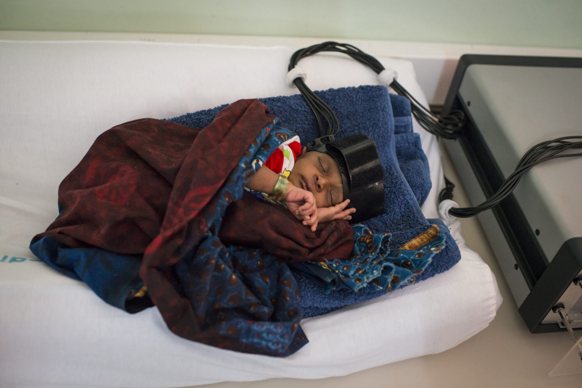
Optical imaging being used to study responses to auditory stimuli in a sleeping newborn infant.
Early assessment of cognitive development may reveal the impact of nutritional deficiencies and allow evaluation of the effectiveness of interventions. Neurobehavioral assessments can be performed in young infants and some have been used to investigate links between nutritional deficit and memory development. However, many of these behavioural tests can only be used to detect effects once they reach the point of observable behaviour, thus reducing the efficacy of early intervention strategies. Furthermore, these assessments are developed based on Western norms, drawing into question their validity outside of these populations. Objective, non-invasive, field-friendly tools for imaging the brains of young infants and children are urgently needed, but the currently employed techniques have limitations for studies of young infants in the developing world. Event related potentials (ERP) can be used to provide objective measures of brain electrical activity but provide only limited spatial localisation. Magnetic resonance imaging techniques (including fMRI) provide a wealth of information on brain structure and function but are restricted to studies in older children and are not suited to studies in the field.

Optical imaging being used to study responses to visual stimuli in a newborn infant.
Near Infrared Spectroscopy (NIRS) is an optical imaging technique, which provides a continuous, non-invasive measure of regional blood flow in the brain. NIRS uses near infrared light which passes through the skull to measure the colour of the blood in the brain. Oxygenated blood appears bright red and is directed to different regions depending on the local brain activity. By using near infrared light to measure the distribution of oxygenated (red) blood we can map brain function. A typical system contains pairs of optical source and detector probes, which are placed on the scalp over regions of interest and are fixed with a lightweight headband that can be adjusting for varying head sizes of infant. Low light levels are used and continuous measurements can be performed with no risk of damage to the tissue. NIRS technology is portable, low cost and requires minimal set up and time and expert training. The technique is completely non invasive and is tolerant of participant motion. For this reason systems have found widespread application amongst researchers interested in infant cognitive function and have been used to study subjects from birth into infancy, childhood and adulthood. One of our team, Dr. Sarah Lloyd-Fox from the Centre for Brain and Cognitive Development, Birkbeck University of London has recently reported the use of NIRS to identify a potential early biomarker of autism in infants as young as 4 months of age.

Optical imaging system and two year old Gambian boy.
Following the award of a Phase One, Grand Challenges Exploration Grant from the Bill and Melinda Gates Foundation, we have recently embarked on a project to determine the role of optical imaging in providing biomarkers of brain development in infants in rural Gambia. In collaboration with Prof. Andrew Prentice and Dr. Sophie Moore at the MRC International Nutrition Group at the London School of Hygiene and Tropical Medicine, in February 2013, we transported a custom built NIRS brain imaging system to the MRC Keneba Field Station in The Gambia. The entire system was carried as excess luggage on the flight and then travelled with us from Banjul on the three hour drive to Keneba. We setup, trained a field worker and ran our first study within 2.5 hours of arrival of the NIRS system. During three visits over the past 10 months our group has been working with Dr. Momodou K. Darboe at MRC Kenebato pilot our NIRS protocols in longitudinal and cross sectional studies infants up to 2 years of age. Our NIRS studies have been focused on determining infants’ responses to auditory social (e.g., laughter) compared to auditory non social (e.g., toy rattles) stimuli, as well as to visual social (human peek-a-boo) compared to visual non social (transport images) stimuli. We have acquired optical brain imaging and anthropometric data in a total of 99 infants ranging in age from 10 days to 24 months and in some of these infants we also have data from behavioural assessments. The data analysed from our first visit in February shows selective cortical activation of Gambian infants to visual-social and vocal stimuli, replicating that observed within similar aged infants in the UK. We are currently analysing data from our subsequent visits and looking at relationships between neuroimaging, growth and behavioural measures.

Behavioural assessment of a young infant.
This Phase One grant has demonstrated the viability of optical brain imaging and behavioural assessment of infants over the first two years of life in a field setting. These studies have provided pilot data for further funding applications to investigate brain development and cognitive function in infants with varying trajectories of growth and nutritional status with the aim of informing early interventions, and we look forward to continuing our collaboration with MRC Keneba.